Figure 3.
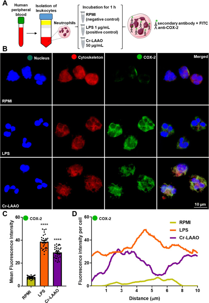
Immunofluorescence of COX-2 in neutrophils. Immunofluorescence of COX-2 using human neutrophils (2 × 10⁵) stimulated Cr-LAAO (50 μg/mL), LPS (1 μg/mL; positive control) or RPMI (negative control) for 1 h at 37 °C and 5% CO2. This figure was created using images from Servier Medical Art Commons Attribution 3.0 Unported License (https://smart.servier.com) (A). Servier Medical Art by Servier is licensed under a Creative Commons Attribution 3.0 Unported License. The images were collected using constant automatic gain among the samples to quantify the differences in absolute levels of fluorescence intensity different conditions in 100 × magnification oil immersion objective. Figure representative of one experiment of three independent experiments (B). Analysis of the mean fluorescence intensity of COX-2 immunofluorescence was performed using 10 cells in field of view of each condition collected impartially (C) and plotted at the fluorescence intensity per cell (D). Values are mean S.E.M. from 3 donors. *P < 0.05, **P < 0.01, ***P < 0.001, ****P < 0.0001 compared to negative control (Data were presented with ANOVA followed by Dunnett post-test).

