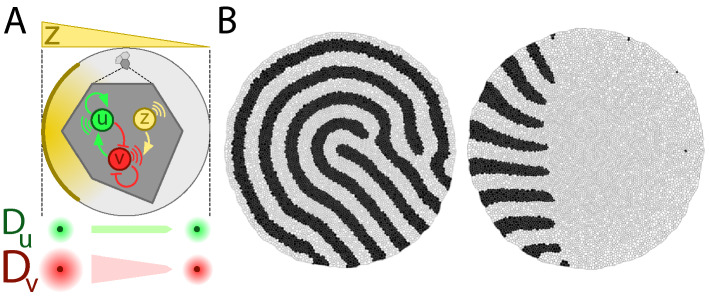
Fig. 4.
Stripe alignment mechanism in growing tissues. (A) In tissues where cells actively grow/divide, if each cell is driven by a regulatory network that just involves an activator, u, and an inhibitor, v, then the resulting Turing pattern displays rotational symmetry (panel B left). If an additional species, z, is released from the “tip” (left side of the tissue in this example) and set a polarity gradient such that the diffusivity of v is spatially modulated, then stripes align following the directionality of the gradient (panel B right). (B) Final snapshot of simulations without (left) and with (right) diffusivity modulation (constant cellular adhesion). The black (white) cellular domains account for regions where (). Since diffusive transport relies on tissue topology (“Methods”) we avoided a possible bias in patterning by using in both simulations the same random sequences that determine the variability of cellular growth/division in order to reproduce the same cellular growth/division events and cell/tissue topologies.

