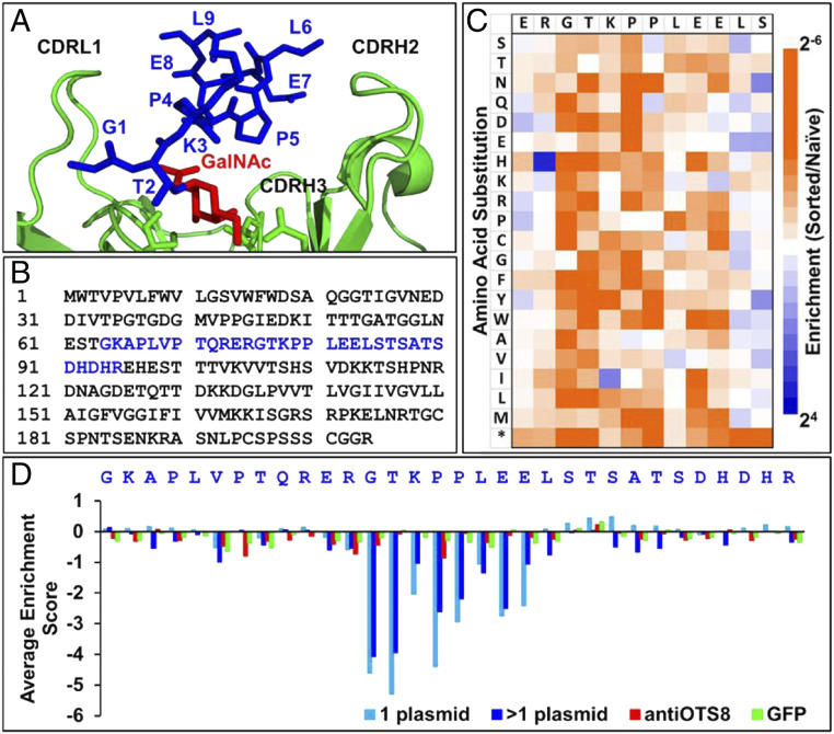
Fig. 1.
Deep mutational scan of the 237-epitope. (A) Binding site of Tn-OTS8 peptide (blue, with GalNAc in red) in 237-monoclonal antibody (green) structure (Protein Data Bank [PDB]: 3IET). (B) Sequence of OTS8 protein, with residues that were subjected to deep mutational scan in blue. (C) Deep mutational scan of OTS8 peptide based on selection of SCLs with 237-IgG. Enrichment or depletion of substitutions was calculated relative to naive (unselected) libraries as a log2 ratio. Resultant enrichment scores were plotted on a color-coded scale ranging from ≤2−6 (orange) to ≥24 (blue). Stop codon is indicated by an *. (D) Average enrichment scores (log2 ratio) for all substitutions in OTS8 SCL when sorted with 237-IgG (blue), anti-OTS8 (red), and GFP (green).

