Fig. 2.
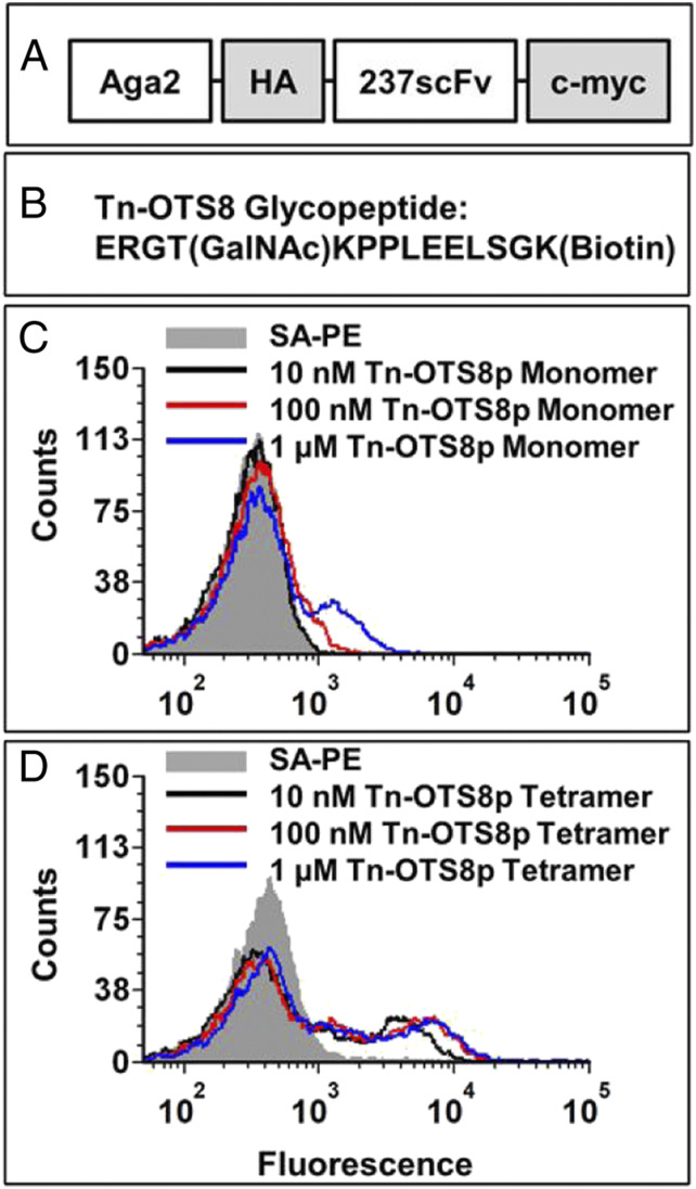
Yeast surface display and antigen binding of 237-scFv. (A) Schematic of 237-scFv cloned in yeast display vector. (B) Sequence of Tn-OTS8 peptide. (C) Yeast-displayed 237-scFv was stained with various concentrations of biotinylated Tn-OTS8 peptide, followed by streptavidin-phycoerythrin (SA-PE). Binding was measured by flow cytometry. The staining profile of yeast cells stained with streptavidin-PE only, is shown in gray. Similar results were obtained in more than three independent experiments. (D) Yeast-displayed 237-scFv was stained with various concentrations of tetramers of Tn-OTS8 peptide made with streptavidin-PE. Binding was measured by flow cytometry. The staining profile of yeast cells stained with streptavidin-PE only, is shown in gray. Similar results were obtained in more than three independent experiments.

