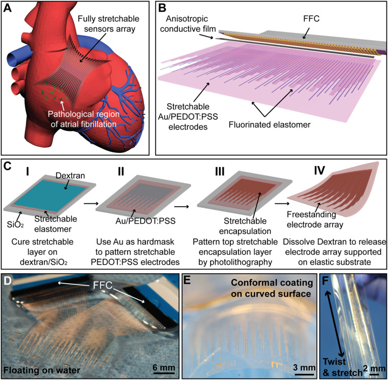
Fig. 1.
Fabrication and assembly of the elastrode array for AF mapping. (A) Schematic showing the application of a fully stretchable elastrode array to the epicardial surface to identify the pathological regions in AF. (B) Schematic showing structure and connections of the elastrode array. (C) Schematics showing the stepwise fabrication processes of freestanding elastrode array. (D–F) Optical photographic images illustrating the ultralight weight (D), high flexibility (E), and stretchability (F) of the freestanding elastrode array.

