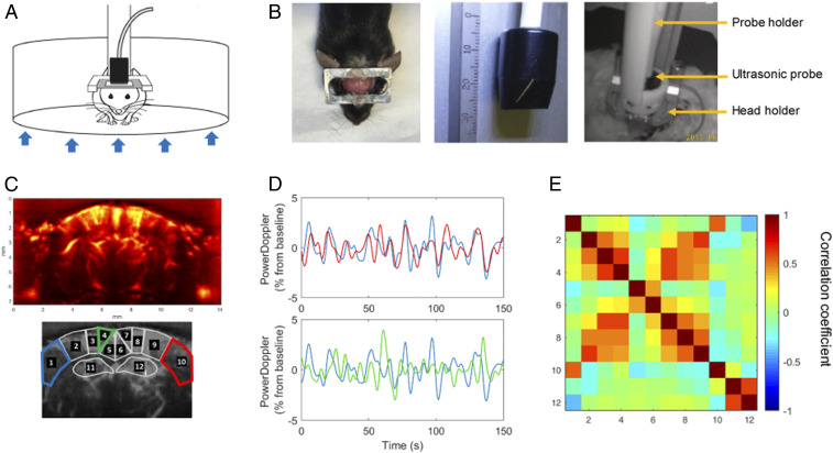
Fig. 1.
fUS in a conscious mouse. (A) Schematic depiction of the experimental set-up for head-fixed freely behaving mice using the air-floated Mobile HomeCage. (B) Mice were implanted with a metal head-frame allowing transcranial ultrasound imaging of a coronal brain section ranging from Bregma to Lambda (Left) with a 15-MHz ultrasonic probe (Center). After application of echographic gel on the imaging window, the ultrasonic probe was lowered at ∼1 mm from the skull using a four-axis motorized probe holder. The mice were held in the dark during the experiments (Right). (C) Representative power Doppler image obtained at approximately −1.5 mm from Bregma. Pixel intensity is directly proportional to CBV. ROIs were selected based on anatomical atlas registration: ROI1 and ROI10: S1BF, primary somatosensory cortex barrel field region; ROI2 and ROI9: S1Tr, primary somatosensory cortex trunk region; ROI3 and ROI8: LPtA/MPtA, parietal associative cortex lateral/median; ROI4 and ROI7: RSA, agranular retrosplenial cortex; ROI5 and ROI6: RSG, granular retrosplenial cortex; ROI11 and ROI12: Hip, hippocampus (dorsal). (D) Normalized average time-series obtained in functionally related regions (1 and 10 corresponding to left and right S1BF cortex, respectively) and unrelated regions (1 and 4 corresponding to left S1BF cortex and left dorsal retrosplenial cortex [RSG], respectively). (E) Resting-state correlation matrix, displaying the mean Pearson correlation coefficient between ROIs defined in C, from a representative awake mouse, showing a 5-min data range from a resting-state epoch (time period without locomotor activity).

