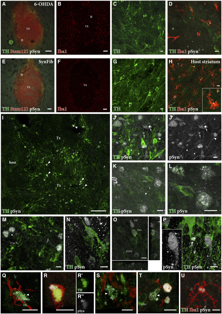
Fig. 6.
pSyn pathology and inflammation in the grafted striatum. Triple staining of TH (green), Stem121 (red), and pSyn (white) is shown in A and E with high magnification of TH+ cells in the regions indicated shown in C and G. Iba1 (red) staining of graft cores is shown in B and F with high-magnification triple staining of TH (green), Iba1 (red), and pSyn (white) shown in D–H. The Inset in H is an Iba1+ cell containing pSyn+ puncta. Double staining of TH (green) and pSyn (white) is shown in I–P with arrowheads in I indicating double-positive cells, which can be seen more easily in high magnification (J–P). Arrowheads in J and J′ indicate pSyn+ deposits were found within (mostly TH-negative) fibers, while arrows in J and J′ indicate pSyn+ deposits extracellularly, outside the TH+ neurons. Double staining within grafted neurons is shown in K–N and P with an orthogonal view in O. Triple staining of TH (green), Iba1 (red), and pSyn (white) in Q–U shows the colocalization of activated microglia around TH+/pSyn+ cells in the graft core. (C) High magnification of area indicated in A; (G) High magnification of area indicated in E; (D) High magnification of area indicated in B. (Scale bars, 100 μm [A, B, E, F, and I] and 10 μm [C, D, G, H, and J–U].)

