Fig. 7.
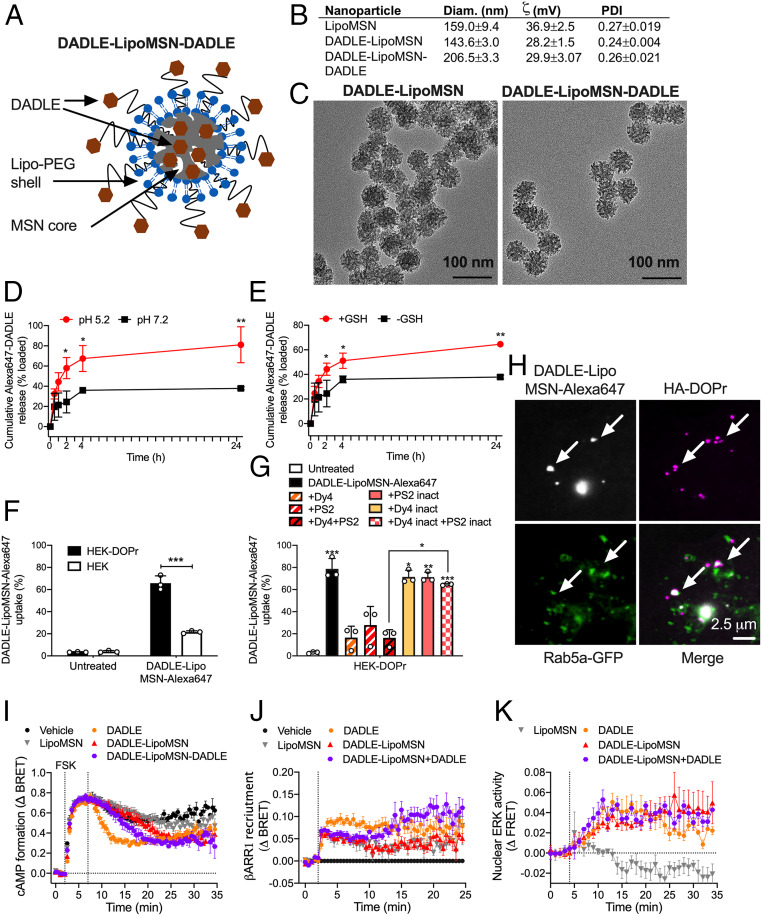
Characterization of nanoparticles. (A) Structure of DADLE-LipoMSN-DADLE. (B) Physical properties of nanoparticles. n = 4 experiments. (C) Transmission electron micrographs of DADLE-LipoMSN and DADLE-LipoMSN-DADLE. Representative images, n = 3 independent experiments. (D and E) Time course of in vitro release of DADLE-Alexa647 from MSN-DADLE-Alexa647 at graded pH (D) and glutathione concentrations (E). n = 3 independent experiments. *P < 0.05, **P < 0.01, t test with Holm–Sidak correction. (F and G) Uptake of DADLE-LipoMSN-DADLE-Alexa647 into HEK293 control and HEK-DOPr cells determined by flow cytometry. (F) Uptake into HEK293 control and HEK-DOPr cells after 2 h. ***P < 0.001, t test with Holm–Sidak correction. (G) Effects of inhibitors of clathrin and dynamin and inactive analogs on uptake into HEK-DOPr cells after 2 h. n = 3 independent experiments. *P < 0.05, **P < 0.01, ***P < 0.001 compared with untreated cells, one-way ANOVA with Tukey’s post hoc test. (H) Uptake of DADLE-LipoMSN-DADLE-Alexa647 into HEK-HA-DOPr cells after 30 min. Arrows show colocalization of DADLE-LipoMSN-DADLE-Alexa647 with DOPr in Rab5a-positive early endosomes. Representative images from four independent experiments. (I–K) Effects of DADLE (100 nM), DADLE-LipoMSN (20 µM), and DADLE-LipoMSN-DADLE (20 µM) on forskolin (FSK; 10 µM)-stimulated cAMP formation (I), βARR1 recruitment (J), and activation of nuclear ERK (K). n = 5 independent experiments. All results are mean ± SEM.

