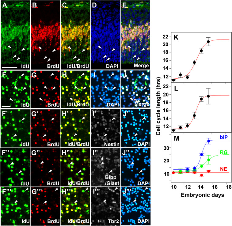
Fig. 2.
Estimation of cell cycle length of cortical progenitors. (A–E) Representative images of E13.5 cortical sections to estimate cell cycle length by the IdU/BrdU dual-labeling method. Embryos were labeled by IdU injection followed by the BrdU 1.5 h later. Embryos were fixed 0.5 h after BrdU injection. S-phase cells in the VZ underneath (labeled by BrdU) were counted as total progenitors. Cells singly labeled by IdU (A and C, arrowheads) and those labeled by BrdU (B) were used for calculating cell cycle length as described in Materials and Methods. (F–J′) Representative images of E13.5 dissociated cortical cells to estimate cell cycle length by the IdU (F)/BrdU (G) dual-labeling method. Total cells were counted as DAPI+ cells (I). (F′–J′′′) E13.5 dissociated cortical cells were stained with IdU (F′–F′′′)/BrdU (G′–G′′′) together with progenitor markers, Nestin (I′), Blbp/Glast (I″) or Tbr2 (I′′′) for the estimation of progenitor type-specific cell cycle lengths. (K–M) Average cell cycle lengths of the entire progenitor population as a function of embryonic days, estimated from IdU/BrdU labeling of cells in sections (K) or dissociated cells (L). Those estimated for each progenitor type are shown in M. (Scale bars: A–E, 50 m; F–J′′′, 50 m.)

