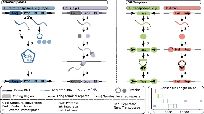
Fig. 1.
TE structure and transposition mechanisms. LTR retrotransposons: 1. Transcription. 2. Translation of one part of the transcripts. The protease (Prot) cleaves pol polyprotein. 3. gag proteins assemble around untranslated transcripts, the integrase (Int), reverse transcriptase (RT) and a tRNA. 4. Reverse transcription and integration. LINE retrotransposons: 1. Transcription. 2. Translation. 3. Protein(s) bind to the transcript. 4. A strand of donor DNA is cut, target-primed reverse transcription starts at the exposed 3’ extremity. 5. The TE is integrated. TIR DNA transposons: 1. Transcription. 2. Translation. 3. Two transposases bind to the TIRs. 4. Transposases dimerize and cut TIR extremities forming a free complex. 5. The complex binds to donor DNA and is integrated. Helitron DNA transposons: 1. Transcription. 2. Translation. 3. At the donor site, the plus strand is cut. A replication fork is formed. 4. Replication results in a double stranded transposon circle. 5. Integration. The bottom right panel represents the distribution of the lengths of D. melanogaster consensus sequences (RepBase [56]), using the same color code as above.

