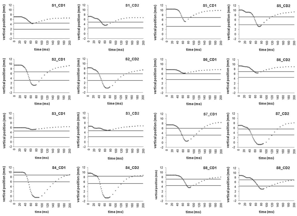
Fig. 1.
Shows as a function of time after TMS the vertical position of the upper eyelid border above the pre-TMS lower eyelid border for different current directions and subjects. This vertical position was measured as the distance between the inner side of the lower border of the upper eyelid after TMS and the inner side of the upper border of the lower eyelid before TMS on a vertical line through the center of the pupil. The upper and lower horizontal lines show, respectively, the position of the upper and lower pupil border before TMS. The Y = 0 coordinate corresponds to the position of the lower eyelid border before TMS. A negative and positive vertical position indicates that the upper eyelid border was, respectively, below and above the pre-TMS lower eyelid border; a decreasing and increasing vertical position indicates that the upper eyelid border is, respectively, descending and ascending. Data are plotted from time of TMS to 200 ms later. The position of the upper eyelid border was measured every 2 ms before its peak descent amplitude and every 10 ms after its peak descent amplitude. a, b Show data for the first and second four subjects, respectively

