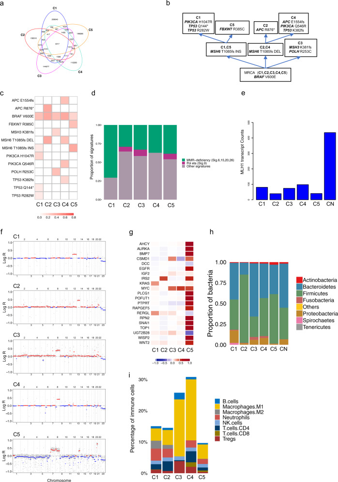
Fig. 3. Genomic and transcriptomic analyses for patient C.
a Venn diagram of SNVs shows low overlap between tumours. b Putative phylogenetic tree based on driver mutations. c VAFs of putative driver mutations. d Mutational signature analysis. e Transcript counts of the MLH1 detected by RNA-seq analysis. f Genomic landscape of CNAs shows low CIN for all samples C1 (ploidy: 2.03), C2 (ploidy: 2.06), C3 (ploidy: 2.13), C4 (ploidy: 2.02) and C5 (ploidy: 2.13). g Median log-ratio of putative driver CNAs highlights heterogeneity of tumourigenic events between all lesions. h Microbial analysis of DNA data shows microbial abundance at the phylum level. i Quantification of tumour immune infiltration reveals varying fractions for eight immune cell populations across all samples.

