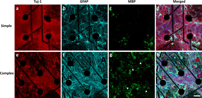
Figure 1.
Immunofluorescence characterization of cortical cultures in simple and complex systems at DIV31. Neurons were identified by staining for Tuj-1 (Neuron-specific class III beta-tubulin, a, e). Glial fibrillary acidic protein (GFAP) was used to identify astrocytes (b, f) and myelin basic protein (MBP) was used to identify mature oligodendrocytes and myelin (white arrowheads) (c, g). Merged images with nuclear stain (DAPI, blue) are shown in (d) and (h). Figure has been modified to remove electrode autofluorescence. Scale bar = 50 µm.

