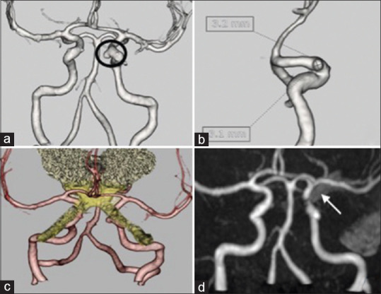
Figure 1.

(a and b) Computed tomography angiogram revealed a saccular aneurysm of size 3.1 mm × 3.2 mm aneurysm in the left internal carotid artery. (c) Fusion image of magnetic resonance imaging with computed tomography angiogram shows the relation of the aneurysm as inferior and closely abutting the medial part of the left optic nerve. (d) Magnetic resonance angiography – postoperative magnetic resonance angiography showing total occlusion of the aneurysm with the aneurysm clip in situ (arrowhead)
