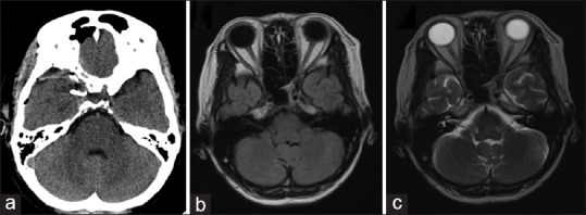
Figure 4.

(a) Computed tomography scan brain plain – postoperative computed tomography scan showing normal brain. Right clinoidectomy can be visualized along with the clip in the right paraclinoid region (white arrow). There was no hematoma visualized in the orbit. (b and c) Magnetic resonance imaging brain plain – postoperative magnetic resonance imaging (a) T1-weighted and (b) T2-weighted images showing a normal study of the optic nerve and the brain
