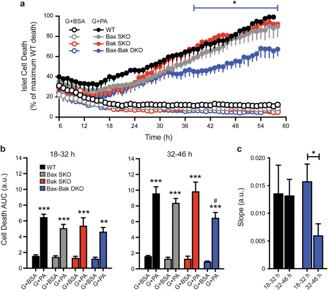
Figure 5.
Combined knockout of Bax and Bak is required for significant protection against glucolipotoxicity. (a) Time-course analysis of cell death (PI incorporation) in cultures of dispersed WT, Bax SKO, Bak SKO, and Bax-Bak DKO islet cells treated with 25 mM glucose + BSA (G + BSA; open circles) or 25 mM glucose + 1.5 mM palmitate (G + PA; closed circles) for 58 h. Between experiments the degree of glucolipotoxic death after 58 h showed some variability (15–25% between islet cell preparations), but was always lowest in the Bax-Bak DKO cells. To facilitate analysis, the islet cell death was expressed relative to that in the WT cultures at the end of each individual experiment (100%). The blue line above the graph indicates the time range where *p < 0.05 for Bax-Bak DKO cell death compared to WT, Bax SKO, and Bak SKO cell death. (b) Area under the curve (AUC) analysis of islet cell death profiles in panel a comparing all genotypes and treatment conditions during two 14 h intervals representing early (18–32 h) and late (32–46 h) stages of cell death. **p < 0.01 and ***p < 0.001 comparing G + PA treatment to their respective G + BSA controls. #p < 0.05 for Bax-Bak DKO G + PA treatment compared to WT G + PA treatment. (c) Rates of WT and Bax-Bak DKO islet cell death were compared by calculating the slope of cell death profiles in panel a for the early (18–32 h) and late (32–46 h) stages. *p < 0.05 (n = 4 independent islet cell preparations). Data represent mean ± SEM, a.u. arbitrary units. Statistical comparisons were done using 2-way ANOVA with Bonferroni post-hoc tests.

