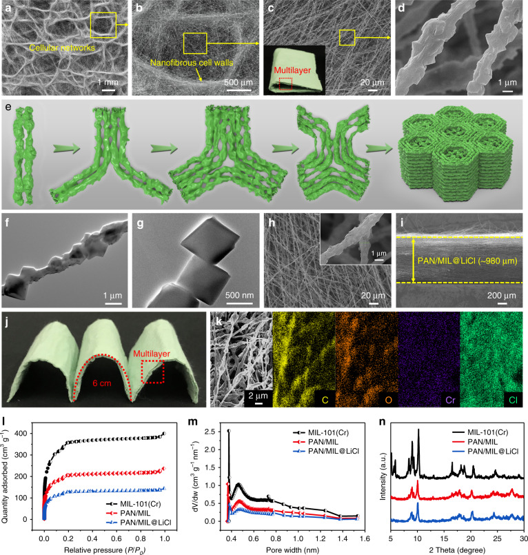
Fig. 2. Morphology and structure characterizations of the desiccant layer.
a–d SEM images at increasing magnifications demonstrating the biomimetic wood-like cellular network structure of PAN/MIL NFM. The inset in (c) is the as-prepared flexible PAN/MIL NFM photograph with a multilayered architecture. e Schematic showing the 3D self-assembly mechanism of PAN/MIL NFM with a multilayer wood-like cellular network structure. TEM images of (f) PAN/MIL nanofiber and (g) pure MIL-101(Cr) crystals. h, i Top-down and cross-section SEM images of PAN/MIL@LiCl NFM. The inset in (h) is a high-magnification SEM image. j Photograph demonstrating the flexibility of PAN/MIL@LiCl NFM and its multilayered architecture. k FE-SEM image and corresponding elemental maps of PAN/MIL@LiCl NFM. l N2 adsorption−desorption isotherms, m pore size distribution curves, and n XRD patterns of MIL-101(Cr) nanoparticles, PAN/MIL, and PAN/MIL@LiCl NFMs, respectively.

