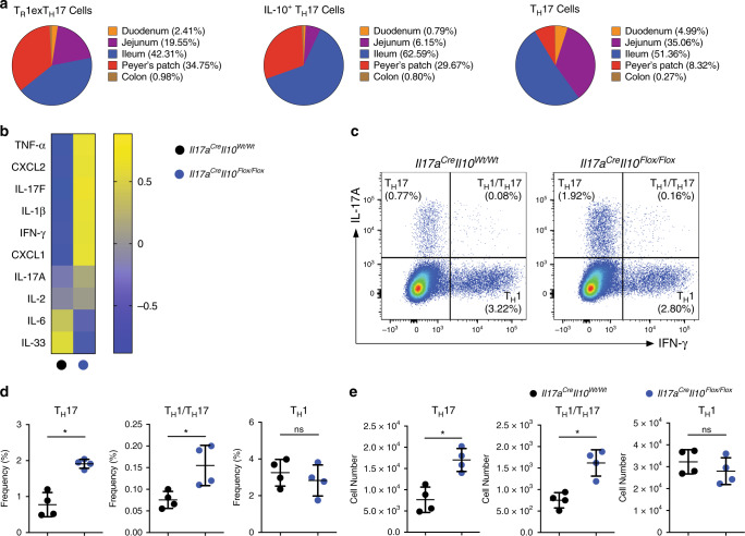
Fig. 1. TH17 derived IL-10 contributes to intestinal homeostasis.
a Tissue distribution of the indicated cell populations within the intestine. Cells were isolated from indicated intestinal tracts of the Fate+ mice. All cell populations are pre-gated on Foxp3−, YFP+, CD4+ T cells and then described as TR1exTH17 (IL-10eGFP+ IL17AKata−), IL-10+ TH17 (IL-10eGFP+ IL17AKata+) and TH17 (IL-10eGFP– IL17AKata+) cells on the basis of the reporter molecules. Cell numbers from three cumulative experiments are used to calculate mean percentage values of the indicated cell populations in different intestinal compartments. b Heatmap showing normalized mRNA expression value (Z-score) of different cytokines/chemokines in small intestinal tissues. c. Flow cytometric analysis of small intestinal CD4+ T cells isolated from the indicated mouse lines under steady state. Intracellular staining for both IL-17A and IFN-γ was then performed to identify TH17 (IL-17A+ IFN-γ−), TH1/TH17 (IL-17A+ IFN-γ+) and TH1 (IL-17A− IFN-γ+) cells. A pre-gate on CD4+ T cells is applied. d, e Statistical analysis of frequencies (d) and numbers (e) are reported. One representative experiment out of three is shown. Each dot represents one mouse (nwild type = 4, nKO = 4). Mean ± S.D.; ns, not significant; *P < 0.05 by Mann–Whitney U test. Source data are provided as a Source data file.

