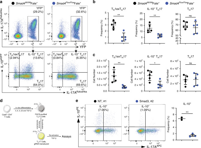
Fig. 5. TGF-β signals via Smad3/4 to regulate IL-10 in TH17 cells.
a Flow cytometric analysis of small intestinal CD4+ T cells after anti-CD3 mAb. The indicated reporter mouse lines were used. Top panels are pre-gated on Foxp3−, CD4+ T cells. Bottom panels are pre-gated on YFP+ cells, and the indicated populations are identified on the basis of the reporter molecules as follows: TR1exTH17 cells: IL-10eGFP+ IL-17aKata−; IL-10+ TH17 cells: IL-10eGFP+ IL-17aKata+; TH17 cells: IL-10eGFP− IL-17aKata+. b, c Frequencies (b) and numbers (c) of TR1exTH17, IL-10+ TH17 and TH17 cells in A are one representative experiment out of three. Each dot represents one mouse (nwild type = 5, nKO = 5). Mean ± S.D.; ns, not significant; *P < 0.05, **P < 0.01 by Mann-–Whitney U test. d Experimental design of knocking out Smad3 in in vitro differentiated mature TH17 cells by using CRISPR/Cas9 technology. e Flow cytometric analysis of in vitro cultured mature TH17 cells after knocking out Smad3 by using gRNAs targeting Smad3. Cells were pre-gated on viable CD4+ T cells. NT, non-targeting control. Each dot represents an individual gRNA (nNT = 2, nSmad3 = 3). Mean ± S.D.; **P < 0.01 by Welch’s t-test. Source data are provided as a Source data file.

