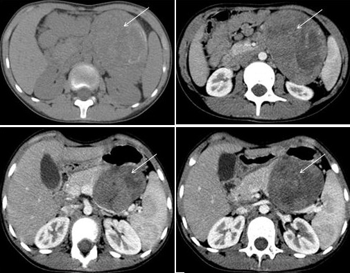
Figure 2.

Computed tomography (CT) imaging identified a well-circumscribed mass located in the body and the tail of the pancreas (arrow). It was heterogeneous, containing cystic and solid portions, with peripheral calcifications. The solid portion had heterogenous enhancement after intravenous contrast administration, demonstrating some necrotic areas
