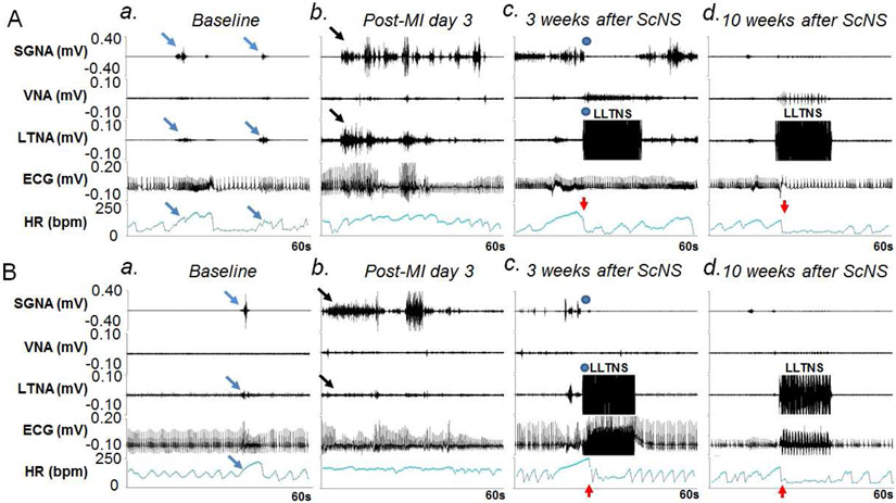
Figure 3.
Effects of ScNS on SGNA, VNA, LTNA and HR. Panels A and B are from two different dogs. Both dogs showed similar patterns of the nerve activity. a: Baseline. Brief nerve activities are associated with HR elevation (arrows). b: Nerve activities (black arrows) significantly increased post-MI, associated with high HR. c: There was an abrupt (blue dots) reduction of SGNA and HR (red arrows) during ScNS ON-time at 3 weeks after the commencement of 3.5-mA ScNS. In Panel Ac but not in Bc was there elevation of VNA during and return of SGNA immediately at the cessation of ScNS. Note that there was increased VNA during the stimulation, along with abrupt reduction of HR (red arrows). d: After 10 weeks of 3.5-mA ScNS, SGNA reduced significantly compared to post-MI period. Onset of ScNS (red arrows) abruptly reduced SGNA, HR and respiratory HR responses. Bradycardia was observed during ScNS ON-time. (ScNS= Left lateral thoracic nerve stimulation, SGNA= stellate ganglion nerve activity, VNA= vagal nerve activity, LTNA= lateral thoracic nerve activity, MI= myocardial infarction).

