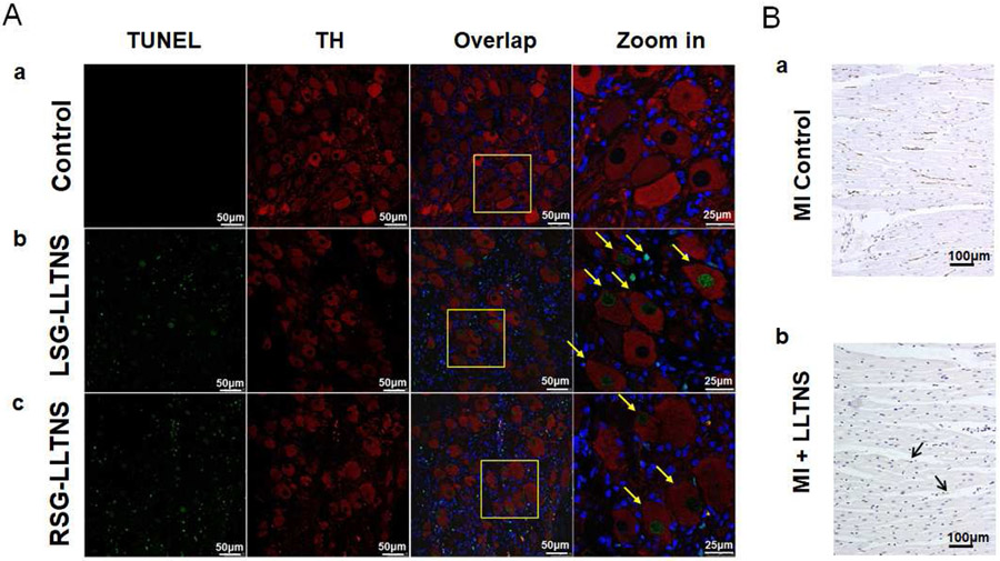
Figure 7.
Effects of ScNS on stellate ganglion and cardiac nerves. A: Confocal microscope images of immunofluorescent Tyrosine hydroxylase (TH, red) and TUNEL (green) double staining in a dog with ScNS. Blue is the DAPI stain of the nuclei. a: Normal stellate ganglion showed no TUNEL positive staining. b and c: Left and right stellate ganglions after ScNS, respectively. Arrows point to ganglion cells that stained positive for TUNEL. Those cells could be either TH-positive or TH-negative (Calibration bar=50 μm). B: Immunostaining of the left ventricle. a. Cardiac nerve sprouting (brown GAP43-positive nerve twigs) are abundantly present in a dog with MI but no ScNS from a previous study.2 In comparison, GAP43-positive nerve fibers (arrows) are rare in the hearts from the present study (b). LLTNS = subcutaneous nerve stimulation using left lateral thoracic nerve. Calibration bar=100 μm.

