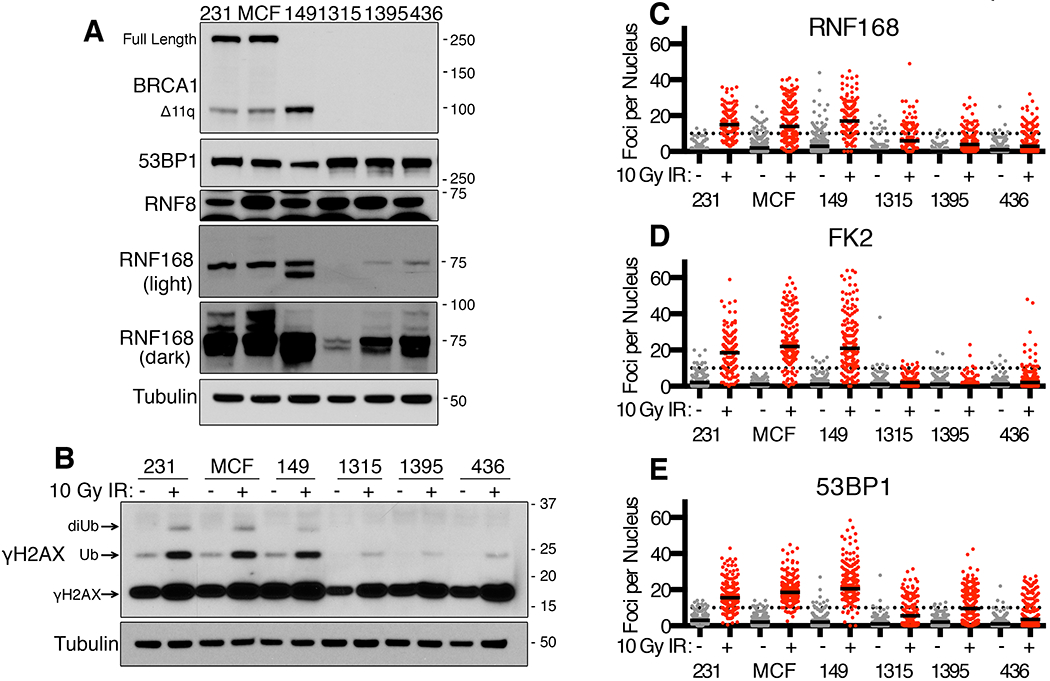
Figure 1. Assessment of RNF168 signaling in BRCA1-deficient cell lines.

(A) MDA-MB-231 (231), MCF7 (MCF), SUM149PT (149), SUM1315MO2 (1315), HCC1395 (1395) and MDA-MB-436 (436) cell lines were assessed for BRCA1, 53BP1, RNF8 and RNF168 protein expression by Western blotting. BRCA1 full length and the BRCA1-Δ11q splice isoform are indicated as well as M.W. markers. Dark and light exposures of RNF168 are shown, also see Fig. S1A.
(B) Western blot analyses of γH2AX in cell lines from (A) at 8h after mock or 10 Gy IR. Mono- and di- ubiquitinated γH2AX forms are indicated.
(C) Cell lines from (A) were analyzed by immunofluorescence (IF) for RNF168 foci at 8h post mock (grey) or 10 Gy IR (red) treatments. The number of foci per nucleus are shown, a minimum of 100 nuclei were assessed. Black solid lines indicate median value; black dotted line indicate 10 foci per nucleus. See Fig. S1B for representative images.
(D) FK2/ubiquitin foci were assessed as described in (C)
(E) 53BP1 foci were assessed as described in (C)
