
Figure 3. Ectopic RNF168 expression decreases the viability of BRCA1 deficient cells.
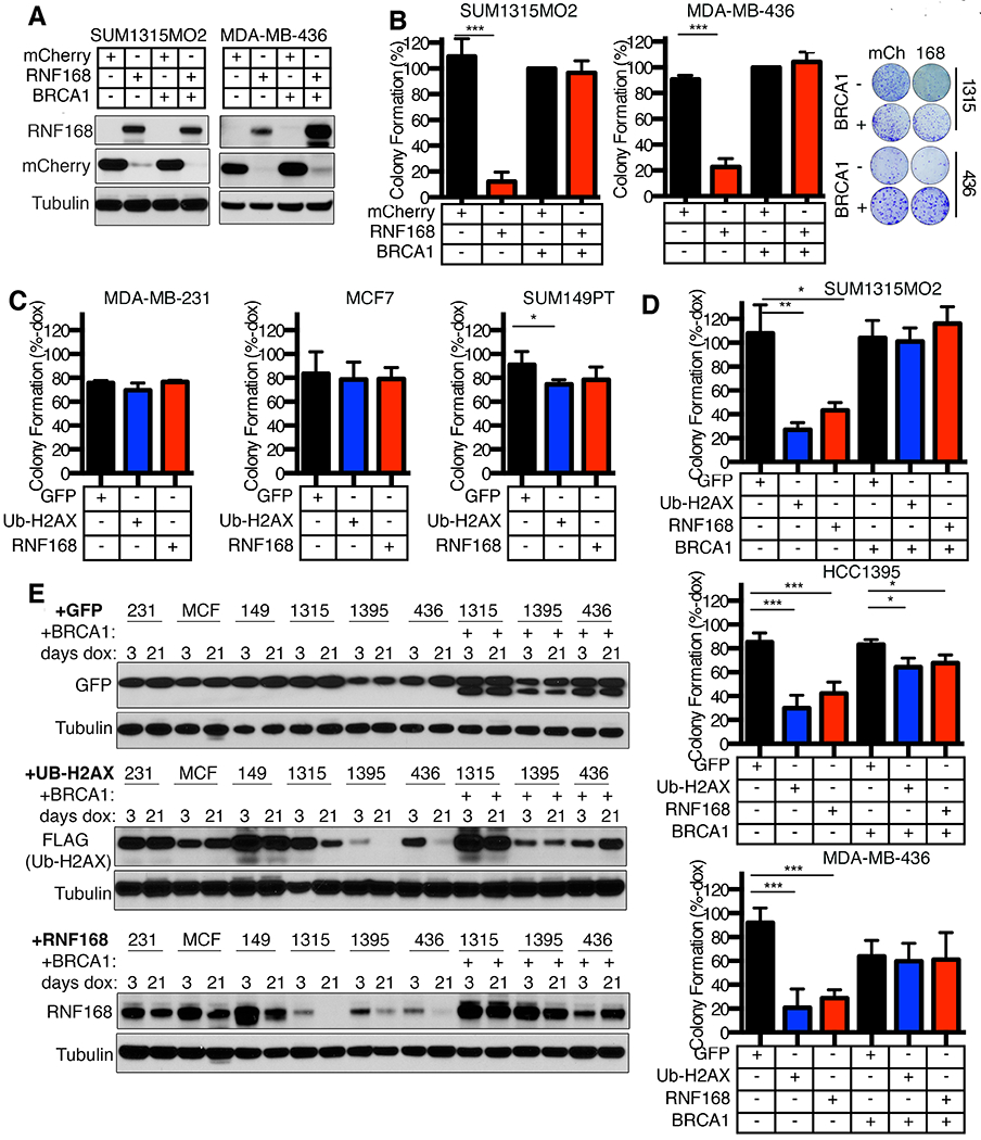
(A) SUM1315MO2 and MDA-MB-436 −/+ BRCA1 add-back cell lines were transduced with lentivirus for expression of mCherry-tagged RNF168 or an mCherry control. mCherry positive cells were sorted and subject to Western blotting for the indicated proteins.
(B) Cells that were mCherry positive from (A) were assessed for colony formation. Colony formation was calculated as a percentage of mCherry expressing BRCA1 add-back cells. Mean ± S.D. is shown, ***p < 0.001 (unpaired t-test). Representative plates and colonies are shown (right).
(C) Dox-inducible GFP, ub-H2AX, or RNF168 expressing MDA-MB-231, MCF7, and SUM149PT cell lines were assessed for colony formation in the absence or presence of dox. Colony formation of dox treated cells is shown as a percentage of colonies formed in the absence of dox. Mean ± S.D. is shown, *p < 0.05 (unpaired t-test). See Fig. S4A for more details.
(D) Dox-inducible GFP, ub-H2AX, or RNF168 expressing SUM1315MO2, HCC1395, and MDA-MB-436 cell lines, as well as isogenic BRCA1 add-back cell lines, were assessed for colony formation in the absence or presence of dox. Colony formation of dox treated cells is shown as a percentage of colonies formed in the absence of dox. Mean ± S.D. is shown, *p < 0.05, **p < 0.01, ***p < 0.001 (unpaired t-test)
(E) Cell lines from (C) and (D) were cultured in the presence of dox for 3 or 21 days and Western blotting performed assessing the indicated proteins.