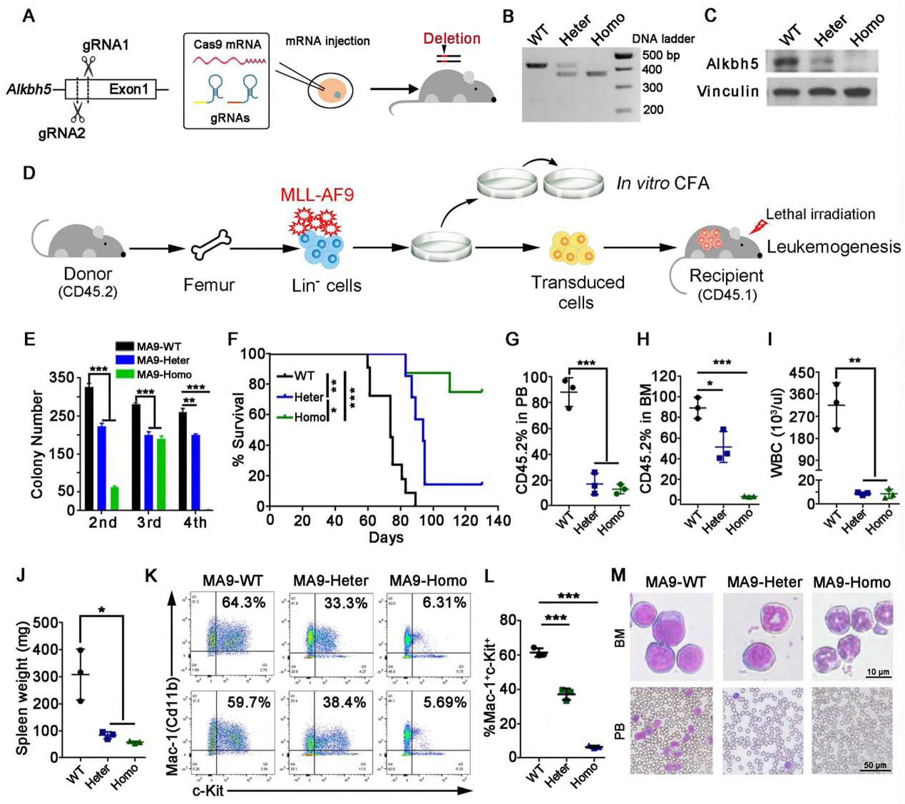
Figure 2. The role of Alkbh5 in MLL-AF9 (MA9) mediated leukemogenesis.

(A) Scheme of the design and procedures of generation of Alkbh5 knockout (KO) mice using the CRISPR-Cas9 technology.
(B and C) Representative DNA genotyping (B) and Western blot assay (C) data of the samples from Alkbh5 wild-type (WT), heterozygous (Heter) or homozygous (Homo) KO mice were shown.
(D) Bone marrow (BM) lineage negative (Lin−) cells collected from Alkbh5 WT, Heter or Homo KO mice were transduced with MA9 retrovirus and used for colony-forming/replating (CFA) assays. The MA9-transduced cells were also transplanted into lethally irradiated recipient mice after first round of colony formation for leukemogenesis.
(E) Colony forming cell counts at each round of plating were shown (n=3).
(F) Kaplan-Meier survival curves of recipient mice transplanted with MA9-transduced Alkbh5 WT (n=11), Heter (n=7) and Homo (n=8) HSPCs.
(G-M) Three mice from each transplant group were euthanized at the same time (day 61 post bone marrow transplantation (BMT)) for AML development analysis. (G-H) Percentage of CD45.2+ cells in peripheral blood (PB) (G) and BM (H) of recipient mice. (I) WBC count in PB of recipient mice. (J) Spleen weight of recipients. (K-L) Percentage of Mac-1+c-Kit+ cells in recipient mice. Representative flow cytometry plots (K) and statistics analysis (L) are shown. (M) Representative images of Wright-Giemsa staining of BM and PB from recipient mice.
*p < 0.05; **p <0.01; ***p < 0.001; t test (for Figures 2E, G–J and L; Mean±SD values are shown) or log-rank test (for Figure 2F). See also Figure S2.
