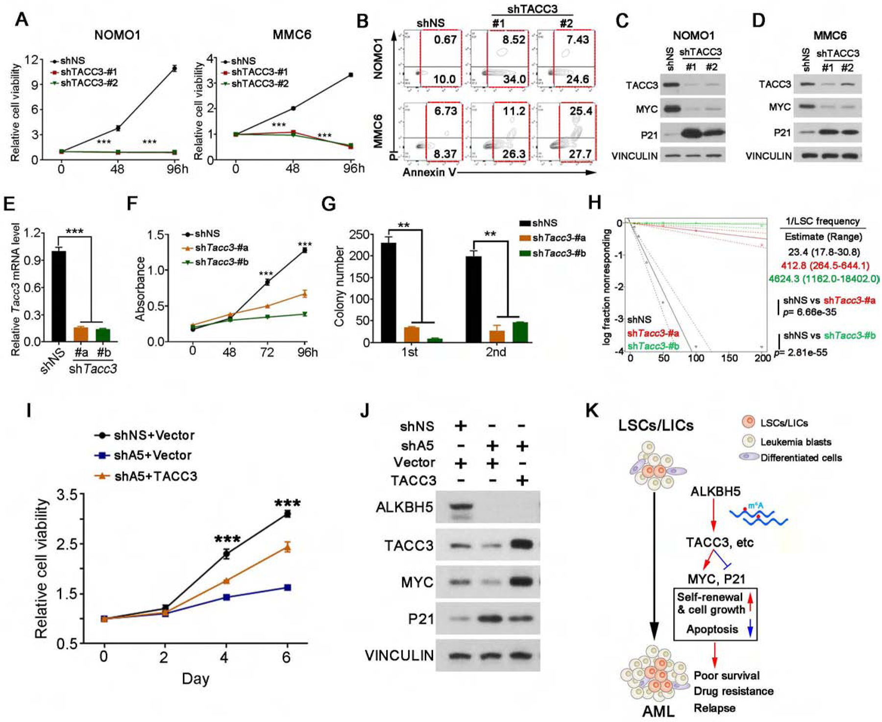
Figure 7. TACC3 is a functionally important target of ALKBH5 in AML.

(A-B) Effects of ALHBK knockdown on cell growth/proliferation assays (A) and apoptosis (B) in AML cells.
(C-D) Western blots of TACC3, MYC and P21 in NOMO1 cells (C) and MMC6 cells (D) expressing shNS or TACC3 shRNAs. VINCULIN was used as a loading control.
(E) Relative levels of Tacc3 mRNA in mouse MA9 AML cells transduced with shNS or individual Tacc3 shRNAs (shTacc3-#a and shTacc3-#b).
(F) Effects of Tacc3 knockdown on the viability/proliferation of mouse MA9 AML cells.
(G) Effects of Tacc3 knockdown on the colony-forming/replating capacity of Mouse MA9 AML cells. Colony forming cell counts at each round of plating are shown (n=3).
(H) In vitro limiting dilution assays (LDAs). Logarithmic plot showing the percentage of non-responding wells at different doses. Non-responding wells, wells not containing colony forming cells. The estimated LSC/LIC frequency is calculated by ELDA and shown on the right. The p value was detected by the log-rank test.
(I-J) MMC6 cells were transduced with shNS or ALKBH5 shRNA (shA5), together with an empty (vector) or TACC3-encoding lentivirus as indicated. After drug selection, those co-transduced cells were seeded into 96-well plates for cell growth/proliferation assays (I). (J) Western blots of ALKBH5, TACC3, MYC and P21. VINCULIN was used as a loading control.
(K) Proposed model demonstrating the role and underlying mechanism(s) of ALKBH5 in AML pathogenesis and LSC/LIC self-renewal.
Mean±SD values are shown for Figures 7A, E–G and I. **p <0.01; ***p<0.001, t test. See also Figure S7.
