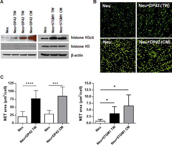
Figure 1. Myeloma cells induce NET formation.
Neutrophils (Neu) were isolated from the BM of tumor-free mice and cultured in vitro alone (Neu), in the presence of mouse myeloma DP42 or 5TGM1 cells separated by Transwell insert (TW), or in the presence of DP42 or 5TGM1 conditioned medium (CM). (A) Western blot analysis of citrullinated histone H3 (H3cit) and total histone H3 in cell lysates prepared from neutrophils cultured for 4h. Experiment was repeated at least three times with similar results. (B,C) NET formation was evaluated using fluorescent microscopy. Representative images of NETs induced by DP42 cells or DP42 cell CM (magnification 20x, scale bar: 50μm) (B) and quantitation of NETs induced by DP42 and 5TGM1 cells (C). Mean and SD values are shown. Each experiment was performed using neutrophils isolated from at least 3 individual mice. * - p<0.05; *** - p=0.0005; **** - p<0.0001 in Student’s t test.

