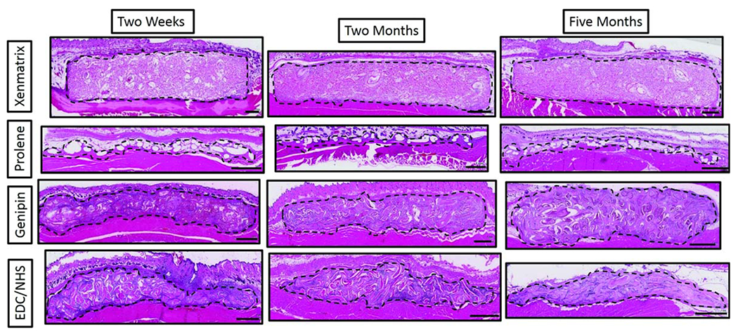
FIGURE 2.

Low magnification light microscopy images of H&E stained mesh sections at 2 weeks, 2 months and 5 months. Meshes were collected along with overlying skin and underlying abdominal muscle. Sections of meshes (within dotted outline) are oriented here with the skin at the top of images and the abdominal muscle at the bottom of the images. Scale bars are 1 mm.
