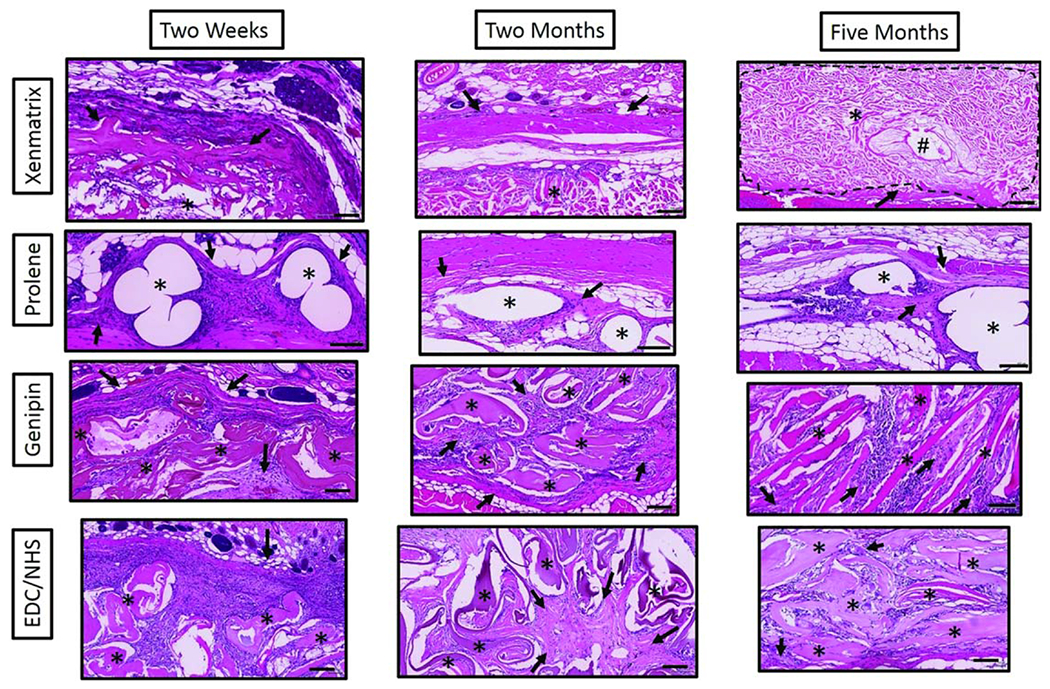
FIGURE 3.

High magnification light microscopy images of H&E stained mesh sections at 2 weeks, 2 months and 5 months. Stars denote the respective mesh materials and arrows indicate areas of new collagen deposition in an around mesh materials. The pound symbol (#) denotes hair follicles in Xenmatrix mesh sections. Scale bars are 100 μm.
