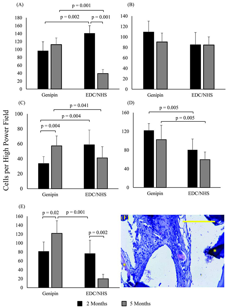
FIGURE 5.

Immunohistochemical identification of macrophage markers in woven collagen scaffolds. (A) General macrophage marker CD68, (B) pro-inflammatory macrophage markers B7, and (C) IL6, anti-inflammatory macrophage markers (D) CD163, and (E) CD206. In all bar graphs n = 15 per time point and horizontal connecting bars indicate a significant difference (p <0.05) between the number of expressive cells for groups. Error bars indicate the SD for test groups (F) Immunohistochemical image of CD206 positive macrophages in genipin crosslinked collagen meshes. Asterisks highlight collagen threads. The scale bar is 0.25 mm.
