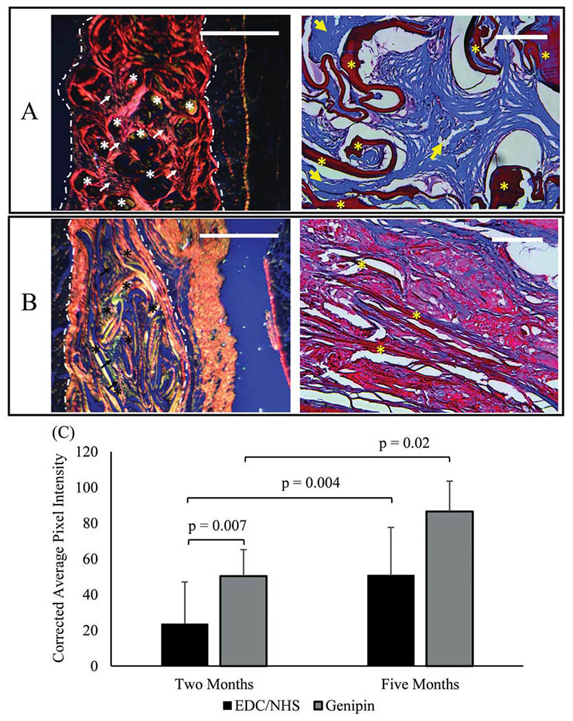
FIGURE 8.

Five months explanted GCC (A) and ECC (B) meshes stained using picrosirius red (left) and Masson’s trichrome (right). Picrosirius red stained slides were imaged under cross polarized conditions. Highly aligned collagen is predominant in GCC samples as manifested by red/orange polarization patterns and loosely aligned collagen is predominant in ECC as manifested by yellow/green polarization in picrosirius stained sections. Masson’s trichrome images show ample amount of de novo collagen deposition (arrows) in genipin crosslinked threads (asterisk). On the other hand, there is minimal collagen deposition in EDC/NHS crosslinked threads. (C) Degree of alignment of newly deposited collagen from picrosirius stained sections was measured by the corrected pixel intensity of images of scaffolds at 2 and 5 months (n = 15 per time point). Significant differences are indicated by connecting bars (p<0.05), and error bars indicate SD for test groups. Scale bars for picrosirius and Masson’s trichrome images are 1 mm and 0.25 mm in length, respectively.
