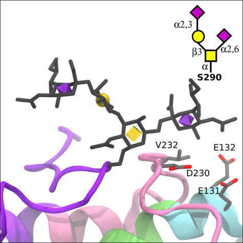
Fig. 6.

Structural changes due to sialylated glycovariants in the lipid-binding domain. The APOE3 crystal structure with a disialylated core 1 glycan (Neu5Acα2–3Galβ1–4[Neu5Acα2–6]GalNAcα) modeled at Ser290 (displayed as licorice with 3D-SNFG icons), which is part of the C-terminal lipid-binding region (purple). Relative to the monosialylated core 1 structure, the α2–6 linked Neu5Ac could potentially form interactions with the C-terminal domain (pink) via V232 and D230, as well as the LDL receptor-binding domain (lime green) via E132 and E131.
