Figure 2.

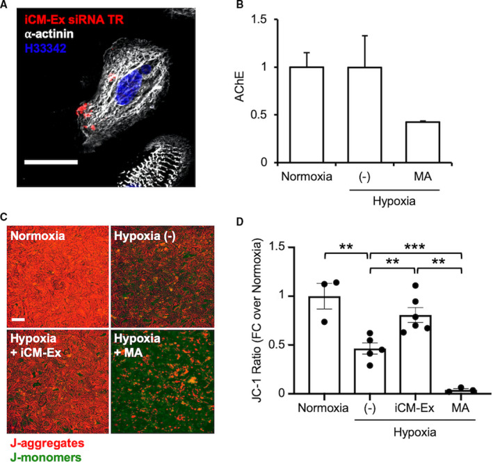
iCM‐Ex are internalized and improve viability in iCMs after hypoxia.
A, iCM‐Ex labeled with fluorescent siRNA are internalized into hypoxic iCMs. Scale bar=50 μm. (C) Supernatants from 3 groups of iCMs were compared: normoxic, hypoxic untreated (−) control, and hypoxic + 10 μmol/L manumycin A (MA). Exosome quantity in the supernatant was measured by acetylcholinesterase (AChE) activity. (C) Mitochondrial membrane potential (MMP) was assessed by JC‐1 fluorescent assay of 4 groups: normoxia, hypoxia untreated (−) control, hypoxia + iCM‐Ex, and hypoxia + 10 μmol/L MA. Scale bar=100 μm. (D) MMP is significantly reduced in hypoxic (−) or MA‐treated cells and restored by iCM‐Ex. **P<0.01, ***P<0.001. FC indicates fold change; iCM, induced cardiomyocyte; iCM‐Ex, exosomes secreted by iCM; siRNA, small interfering RNA; and (−), untreated control.
