Figure 4.

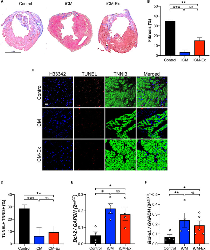
iCM‐Ex improve cardiac cell survival.
A, Masson trichrome–stained mid‐LV sections of saline control, iCM, and iCM‐Ex mice. B, Percentage of fibrotic area in LV. C, LV sections from saline control, iCM, and iCM‐Ex mice were costained with TUNEL (red) and TNNI3 (green) antibodies and Hoecsht 33 342 (blue). Scale bar=20 μm. D, Percentage of apoptotic cardiomyocytes. E, PIR of saline control, iCM, and iCM‐Ex mice was isolated for qRT‐PCR of (E) BCL‐xL and (F) BCL‐2 expression, normalized to GAPDH. Data are mean±SEM of minimum n=3. *P<0.05, **P<0.01, ***P<0.001. iCM indicates induced cardiomyocyte; iCM‐Ex, exosomes secreted by iCM; LV, left ventricular; NS, not statistically significant; PIR, peri‐infarct region; qRT‐PCR, quantitative real‐time polymerase chain reaction; and TUNEL, terminal deoxynucleotidyl transferase dUTP nick end labeling.
