Figure 4.

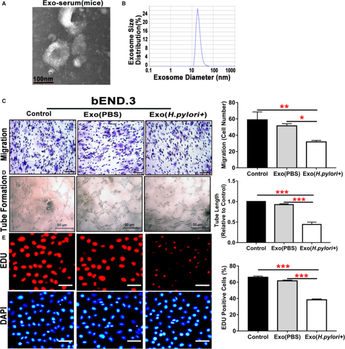
Serum exosomes from mice with Helicobacter pylori infection inhibited endothelial function in vitro.
Exosomes isolated from mice with CagA+ H pylori infection exhibited typical exosome characteristics with unique morphologies as shown on transmission electron microscopy (A), and size distribution as demonstrated using a Zetasizer Nano ZS (Malvern Instruments, Malvern, Worcestershire, UK) instrument (B). Transwell culture showed that exposure to the serum exosomes from mice with CagA+ H pylori infection significantly inhibited the migration of mouse brain microvascular endothelial cells bEND.3 (C, scale bar=50 μm) as compared with the controls. Culture with the serum exosomes from mice with CagA+ H pylori infection also significantly attenuated tube formation (D, scale bar=50 μm) and proliferation (using EdU assay) (E, scale bar=50 μm) of bEND.3. bEND.3 indicates mouse brain microvascular endothelial cells; Exo (H pylori +), xosomes derived from mice treated with CagA+ H pylori infection; Exo (PBS), exosomes derived from control mice treated with PBS; Exo‐serum, exosomes isolated from mouse serum. *P<0.05, **P<0.01, ***P<0.001 by Kruskal‐Wallis test with Dunn post hoc test or 1‐way ANOVA with Bonferroni test. Data were presented as mean±SE, n=3 independent experiments (experiment was repeated 3 times for every measurement).
