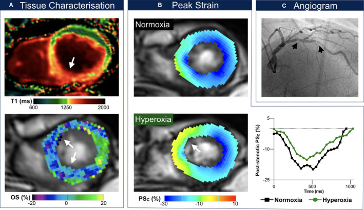
Figure 2.

Exemplary patient with systolic dysfunction. Findings in an 80‐year‐old male patient with serial left anterior descending artery stenoses (C, maximal degree: 63% diameter stenosis). This patient had a previous percutaneous coronary intervention of serial right coronary artery stenoses following presentation with typical stable angina and inconclusive ergometry. In this patient, left ventricular ejection fraction dropped from 64% to 57% and cardiac index from 2.77 to 2.37 L/min/m2 after breathing oxygen, respectively. The T1 mapping image (A, top) shows myocardial injury in the septum and inferior wall, which is colocalized with both the oxygenation deficit (A, bottom) and circumferential strain (PSC) abnormalities induced with hyperoxia. At rest, the septum exhibits subnormal peak circumferential strain at end‐systole (B), which is exacerbated further at hyperoxia (yellow region). The graph shows that peak circumferential strain in poststenotic segments is attenuated across the entire cardiac cycle at hyperoxia (green) in comparison with normoxemia at room air (black). OS (%) indicates % change in oxygenation‐sensitive signal intensity.
