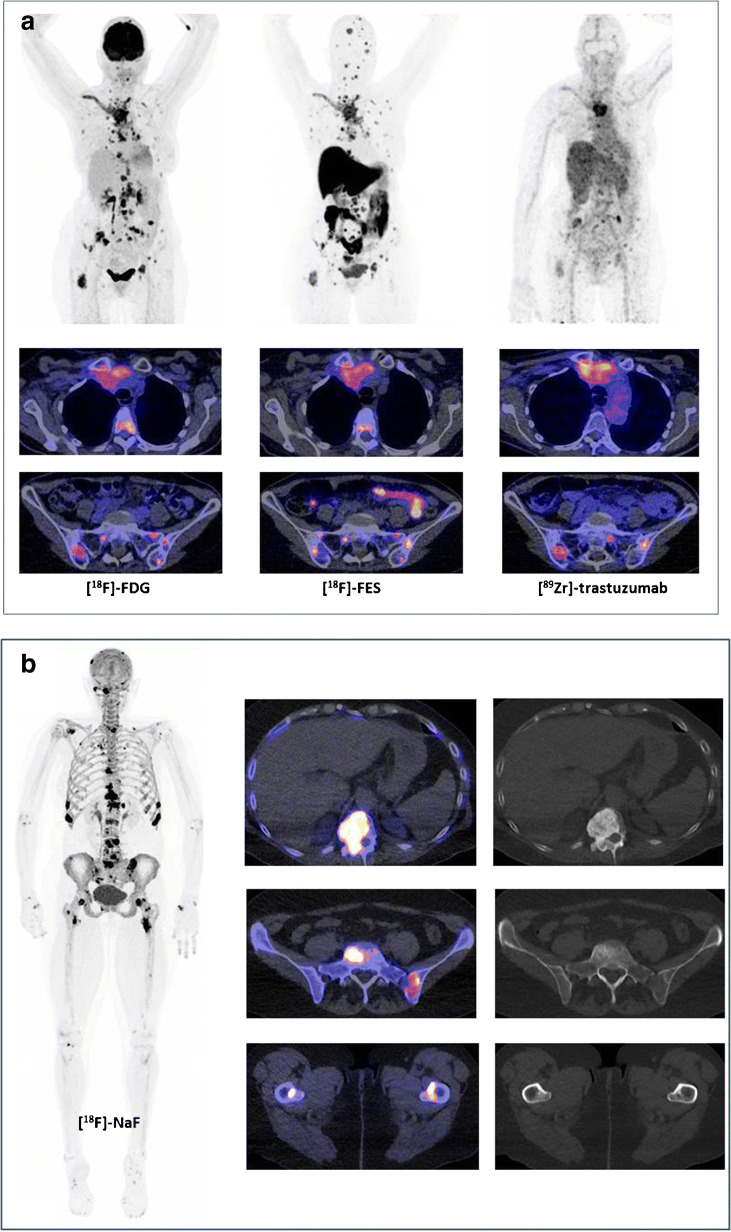
Fig. 1.
Upper image: three PET scans ([18F]-FDG-PET, [18F]-FES-PET, and [89Zr]-trastuzumab-PET) in the same patient showing mediastinal and hilar lymph node metastases, as well as intrapulmonary lesions visible on both [18F]-FDG-PET and [18F]-FES-PET, but not on [89Zr]-trastuzumab-PET. The large mediastinal mass (first row of transversal fused images) was visible on all three imaging modalities. Bone metastases (second row of transversal fused images) were clearly visualized on [18F]-FES-PET, for example, skull lesions, and to a lesser extent on [18F]-FDG-PET and [89Zr]-trastuzumab-PET. Lower image: [18F]-NaF-PET in another patient showing bone metastases in the skull, vertebrae, costae, pelvis, and proximal femora. The increased uptake in the joint was related to degeneration

