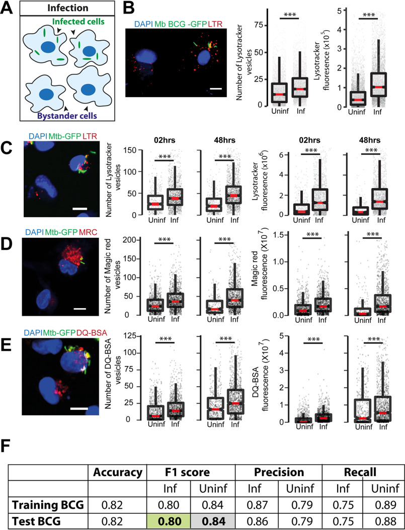
Figure 1.
M. tuberculosis infected macrophages have higher lysosomal content and activity (in vitro). A, schematic showing the experimental design to differentiate between infected and uninfected (bystander) cells in the same population. B, primary human macrophages infected with GFP expressing M. bovis BCG were pulsed with LysoTracker Red at 48 hpi. The number of LysoTracker vesicles and integrated LysoTracker fluorescence intensity were compared between infected and bystander-uninfected cells. C–E, THP-1 monocyte-derived macrophages were infected with M. tuberculosis-GFP H37Rv and pulsed with LysoTracker Red (C), MRC (D), or DQ-BSA (E) 2 and 48 hpi and imaged. Graphs show the number and total cellular intensities of the corresponding vesicles at the indicated time points. Results are representative of at least three biological experiments. Statistical significance was assessed using Mann-Whitney test, *** denotes p value less than 0.001. Scale bar is 10 μm. For B–E, data are represented as box plots, with median value highlighted by a red line. Individual data points corresponding to single cells are overlaid on the boxplots. F, multiparametric data from different infection experiments were used to train a classifier to predict infected cells based on the lysosomal features, as described under “Experimental procedures.” The test was done in the absence of information on the bacteria channel. The close match in the F1 score between training and test datasets indicates accurate prediction. Approximately 15,000 cells were used for the M. bovis BCG training data set, and 6500 for the test.

