Figure 9.
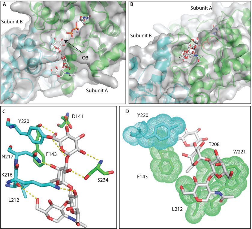
Computational docking explains specificity of Gat1 for the Skp1-tetrasaccharide. A, top docking pose of the Skp1-tetrasaccharide in the PuGat1 active site and a groove formed by the dimer. B, 90° turn of the image shown in A. C, hydrogen-bonding interactions of the glycan. D, hydrophobic packing of the sugar faces and fucose-methyl (in sticks) with nonpolar surfaces (sticks/dots) of Gat1 subunit A (green) and B (cyan).

