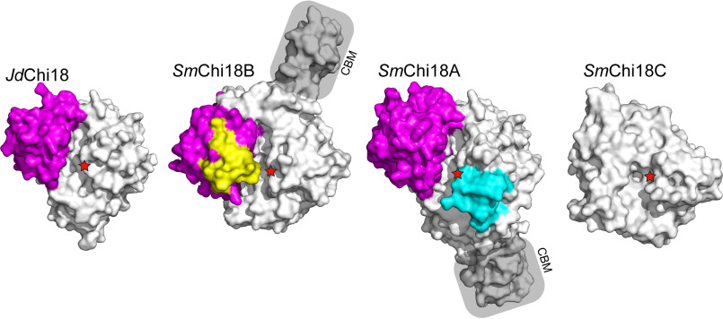
Figure 2.
Structural comparison of JdChi18 with well-studied chitinases from S. marcescens. The figure shows surface representations of the four enzymes, looking into the substrate-binding clefts of the enzymes from the top. The so-called α + β domain is colored magenta, whereas extra loops (relative to JdChi18) in SmChi18B and SmChi18A are shown in yellow and cyan, respectively. The catalytic acid is indicated by red stars in all four enzymes. CBMs are labeled and shaded. Note that for SmChi18C only the catalytic domain is shown and that this domain lacks the α + β insertion. SmChi18C has two additional domains likely involved in substrate binding that are not shown because the structure of the full-length protein is not known.

