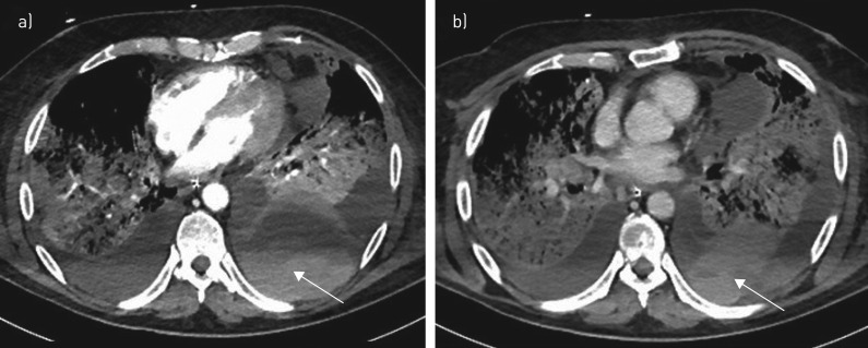
FIGURE 1.
Computed tomography images of a 68-year-old patient with severe acute respiratory distress syndrome-coronavirus-2. Axial images in a) the arterial phase and b) after 80 s show major left circumferential haemothorax with fresh blood (arrows) without active bleeding, complicating lower lobar pulmonary necrosis.

