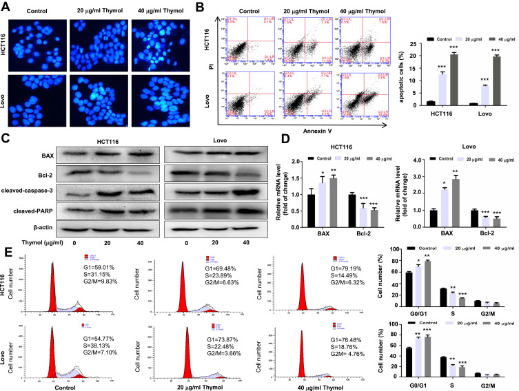
Figure 2.
Thymol induces apoptosis and cell cycle arrest in colorectal cancer (CRC) cells. (A) Effects of thymol on the nuclear morphology of CRC cells. Cells were treated with thymol at different concentrations (0, 20, or 40 µg/mL). DMSO (0.1%) was added as a control. After 48 h, the cells were fixed and stained with Hoechst 33258. The morphology of the cancer cells was observed under a fluorescence microscope (×200). (B) Flow cytometric analysis of apoptosis in CRC cells. Thymol treatment promoted cell apoptosis in a dose-dependent manner. ***P < 0.001 vs the control group. (C) HCT116 and Lovo cells were treated with thymol at different concentrations (0, 20, or 40 µg/mL) for 48 h. DMSO (0.1%) was added as a control. Bcl-2, BAX, cleaved caspase-3, and cleaved PARP protein expression was evaluated by Western blot. (D) The mRNA levels of BAX and Bcl-2 were evaluated by RT-qPCR. GAPDH was used as an internal control. *P < 0.05, **P < 0.01, ***P < 0.001 vs the control group. (E) Cell cycle analysis by flow cytometry. Cells are treated with thymol at different concentrations (0, 20, or 40 µg/mL). DMSO (0.1%) was added as a control. Thymol treatment shortened the S phase and prolonged the G1 phase in HCT116 and Lovo cells. *P<0.05, **P<0.01, ***P<0.001 vs the control group. All data are presented as mean ± SD of three independent experiments.

