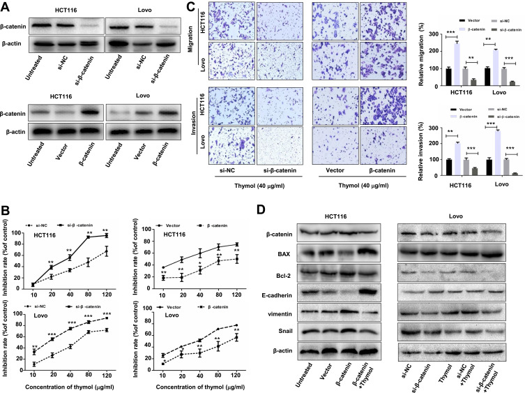
Figure 6.
Thymol inhibits colorectal cancer (CRC) cell proliferation, migration, and invasion by suppressing Wnt/β-catenin signaling. HCT116 and Lovo cells were transfected with β-catenin siRNA (si-β-catenin)/negative control siRNA (si-NC) or the pcDNA3.1-β-catenin plasmid (β-catenin)/pcDNA3.1 (vector). (A) Western blot analysis of β-catenin expression in HCT116 and Lovo cells after transfection with β-catenin siRNA, pcDNA3.1-β-catenin, or the corresponding controls for 24 h. (B) At 24 h post transfection, the cells were exposed to different concentrations of thymol (0, 10, 20, 40, 80, or 120 µg/mL) and cell proliferation was quantified at 48 h. *P<0.05, **P<0.01, ***P<0.001 vs the control group. (C) At 24 h post transfection, cells were plated into the upper chamber of a transwell plate and treated with thymol (40 μg/mL). After 24 h of incubation, the migratory and invasive abilities were assessed. Cells were stained and imaged under a microscope (×100 magnification). **P<0.01, ***P<0.001 vs the control group. (D) At 24 h post transfection, cells were treated with thymol (40 μg/mL) for 48 h. The expression levels of β-catenin, BAX, Bcl-2, E-cadherin, vimentin, and Snail were measured by Western blotting, and normalized to β-actin expression. All data are presented as mean ± SD of three independent experiments.

