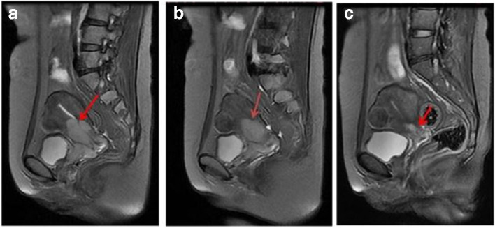
Figure 3.
Difussion-weighted MRI of cervical squamous cell carcinoma These are sagittal T2 weighted images of a 45-year-old patient before treatment (A),15 days after treatment (B), and 2 months after treatment (c). The tumor (indicated by the arrow) size reduced (and the ADC values increased) after treatment. Reproduced from: Chen J. The Value of DW MRI in predicting the Efficacy of Radiation and Chemotherapy in Cervical Cancer. Open Life Sciences. 2018; 13(1) 305-311

