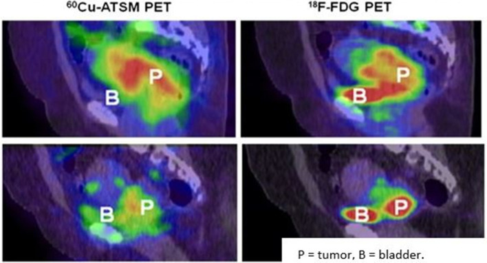
Figure 4.
PET/CT imaging of Cu-ATSM and F-FDG uptake in hypoxic and normoxic cervix tumors Top: These sagittal images are of a patient with a hypoxic cervix tumor. They show high 60Cu-ATSM(left) and high 18F-FDG(right)uptake in the tumor.Bottom: These sagittal images are of a patients with an overall normoxic cervix tumor. There is a sharp decrease in 60Cu-ATSM(left) uptake compared to the hypoxic tumor, but still a fairly high 18F-FDG(right)uptake compared to the hypoxictumors than 18F-FDG.Reproduced from: Lyng H. Hypoxia in cervical cancer: from biology to imaging. Clin Transl Imaging. 2017; 5(4): 373-388.

