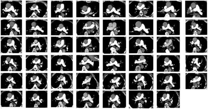
Figure 3.
Axial slices at the level of the main pulmonary artery in all 47 patients included in the test cohort (Phase II). The image quality concerning PE was, by all three readers, visually scored as adequate or better in 47/47 patients (100%, 95% confidence interval 92–100%). PE, pulmonary embolism.

