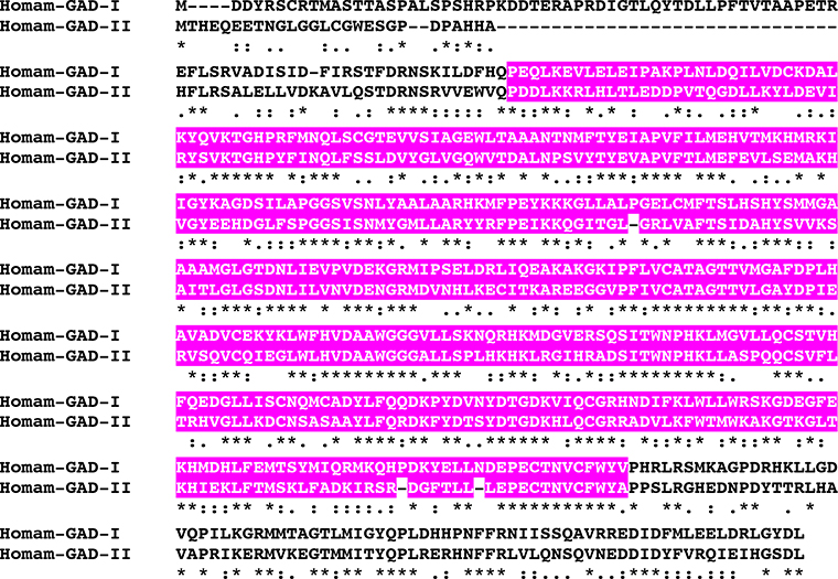
Figure 11.
MAFFT alignment of Homarus americanus glutamic acid decarboxylase I (Homam-GAD-I) and glutamic acid decarboxylase II (Homam-GAD-II). In the line immediately below each sequence grouping, “*” indicates identical amino acid residues, while “:” and “.” denote amino acids that are similar in structure between sequences. In this figure, pyridoxal-dependent decarboxylase conserved domains identified by Pfam analyses are highlighted in pink.

