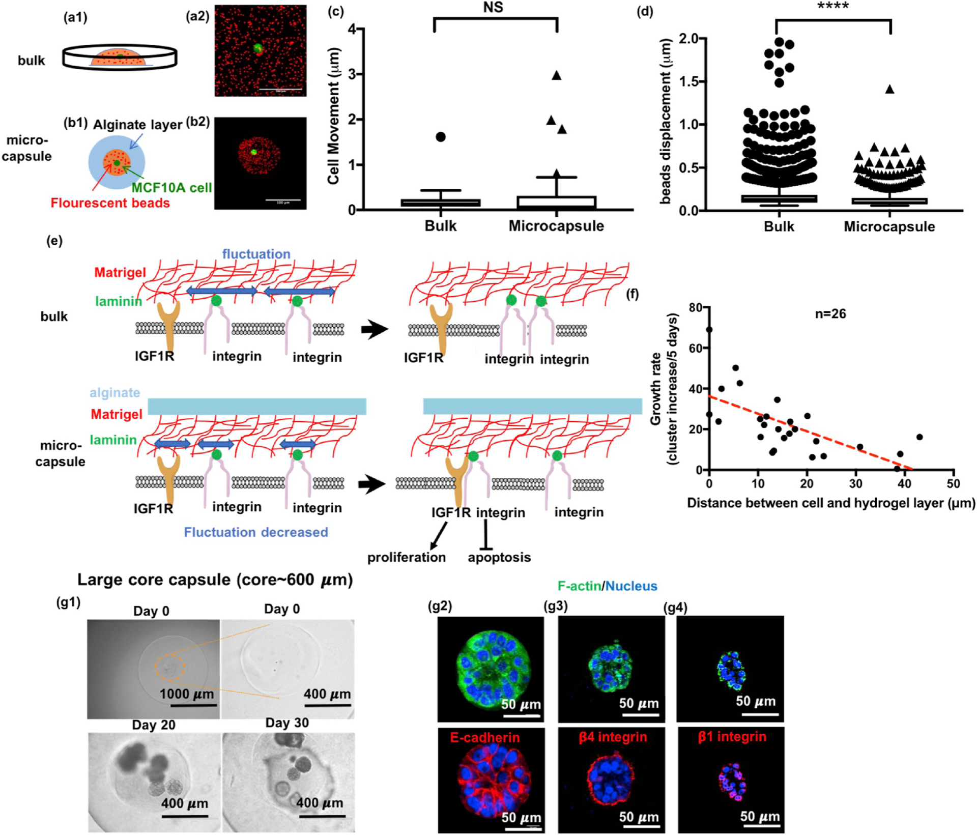
Fig. 5.

Confinement restricts matrix fluctuation to disrupt acini formation. (a-d) Study of matrix fluctuation using fluorescent beads tracking. (a) Schematic and the z-projection of fluorescent beads and a MCF10A cell in bulk Matrigel. (Red: fluorescent beads; Green: GFP-expressing MCF10A cells.) (b) Schematic and z-projection for a single MCF10A cell in Matrigel with fluorescent beads encapsulated in an alginate core-shell microcapsule. Scale bars are 100 μm. (c) Cells behaved similarly in bulk as in microcapsules. (n = 40, 56 pooled from 5 independent experiments, t = 4.75, df = 870.7, p = 0.2993 by Welch-corrected t-test) (d) Bead displacement was significantly smaller in microcapsules than in bulk where beads were freely moved without confinement. (n = 8113, 732 pooled from 5 experiments, t = 1.044, df = 84.6, p < 0.0001 by Welch-corrected t-test) (e) Schematic illustration for how confinement might disrupt the formation of hemidesmosomes while promoting the integrin binding to insulin-like growth factor receptor to induce uncontrolled growth. (f) The relationship for growth rate versus the distance between the cell and the alginate layer. (g) When the core size increased to ~ 600 μm, the confinement effect diminished. The confined cells formed acini again in these capsules, integrin β1 (red) and β4 (red) were localized on the surface and E-cadherin (red) was fully expressed.
