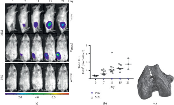
Figure 1.

Detection of MOPC315.BM.Luc cells in 26-week-old BALB/c mice following inoculation. (a) BLI images of ventral and lateral views of a MOPC315.BM.Luc- and a PBS-injected mouse at increasing days after inoculation. (b) BLI signals (total flux in photons−1) of one control and ten MM mice at days after inoculation (median, interquartile range). (c) Typical femoral injection site (white arrow) in a 3D microCT reconstruction at day 21.
