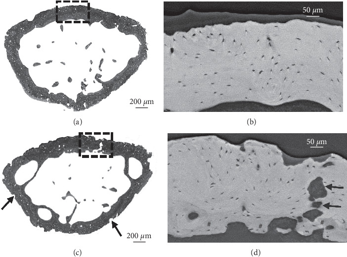
Figure 4.

PCE-CT for bone ultrastructural characterization of femora injected with PBS (a, b) versus MOPC315.BM.Luc cells (c, d) at day 21. (a, c) PCE-CT images of cross sections of femur metaphysis. (c) Numerous sites of erosion (black arrows) and bone loss are indicated in the femur of the MM-injected bone. (b, d) High-magnification view of the rectangular area in (a) and (c). (d) Rectangular area of a local region in the cortex reveals multiple zones of intensive bone remodeling activity, with low-density new bone (darker grey, indicated with #) intermixed with mature bone sites (brighter grey), pocked with irregular-shaped “punched-out” lesions (black arrows).
